Home > Arts > Artists > J > Jacob Jacobs
Heart and lungs

![]()
Wall Art and Photo Gifts from Science Photo Library
Heart and lungs
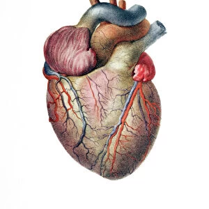
Heart and lungs. Historical anatomical artwork of the human heart and lungs, seen from the front. Dissection hooks have been used to draw back the lungs (red, left and right) to reveal the heart (centre) with its pericardium partially opened to reveal the blood vessels on the surface of the heart. At upper centre the trachea (windpipe, white) supplies air to the lungs. Either side of the trachea are the major blood vessels (carotid arteries, red; jugular veins, blue) of the neck. The major shoulder blood vessels are also seen. Artwork from the 19th-century book Atlas of Anatomy, by Bourgery and Jacob. This book, which took over 20 years to complete, was published in France in 8 volumes from 1831 to 1854. It contained 726 colour plates covering both anatomy and surgical techniques
Science Photo Library features Science and Medical images including photos and illustrations
Media ID 1700375
© MEHAU KULYK/SCIENCE PHOTO LIBRARY
Anatomical Artwork Anterior Blood Circulation Cardiac Cardiology Chest Dissected Dissection Drawing French Frontal Jacob Jean Baptiste Marc Bourgery Lungs Neck Nicolas Henri Jacob Pulmonary Respiratory System Thoracic Thorax Trachea Circulatory System
FEATURES IN THESE COLLECTIONS
> Arts
> Artists
> J
> Jacob Jacobs
> Science Photo Library
> History
EDITORS COMMENTS
This 19th-century anatomical artwork showcases the intricate beauty of the human heart and lungs. In this print, we are presented with a frontal view of the chest, where dissection hooks have delicately pulled back the vibrant red lungs to unveil the central masterpiece - the heart. The pericardium has been partially opened, allowing us a glimpse into its inner workings and revealing the intricate network of blood vessels that adorn its surface. At the upper center of this illustration, we can observe the trachea or windpipe in pristine white supplying air to both lungs. Flanking either side of it are prominent blood vessels - carotid arteries in striking red and jugular veins in serene blue - which serve as vital conduits for circulation within our necks. Additionally, we catch sight of major shoulder blood vessels further enriching this comprehensive depiction. This remarkable artwork is an excerpt from "Atlas of Anatomy" an extraordinary French publication by Bourgery and Jacob that spanned over two decades to complete. Comprising eight volumes released between 1831 and 1854, this monumental work encompassed 726 color plates illustrating not only anatomy but also surgical techniques. Through this historical image, we gain insight into centuries-old medical knowledge while marveling at the complexity and elegance inherent within our own bodies' circulatory system. It serves as a testament to humanity's unending quest for understanding our biological selves while paying homage to pioneers like Jean Baptiste Marc Bourgery and Nicolas Henri Jacob who dedicated their lives to advancing medical science through artistry.
MADE IN THE USA
Safe Shipping with 30 Day Money Back Guarantee
FREE PERSONALISATION*
We are proud to offer a range of customisation features including Personalised Captions, Color Filters and Picture Zoom Tools
SECURE PAYMENTS
We happily accept a wide range of payment options so you can pay for the things you need in the way that is most convenient for you
* Options may vary by product and licensing agreement. Zoomed Pictures can be adjusted in the Cart.