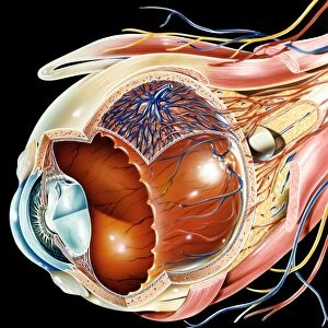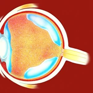
Eye anatomy, artwork

![]()
Wall Art and Photo Gifts from Science Photo Library
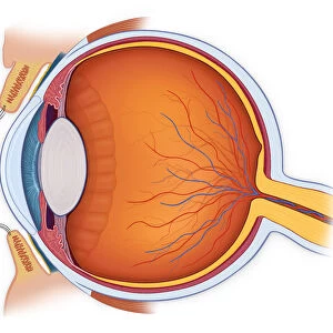
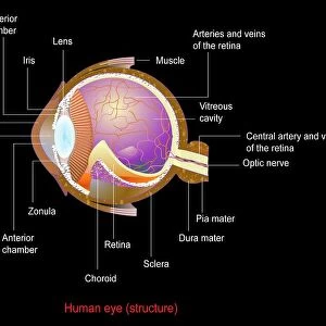
Eye anatomy, artwork
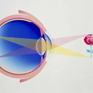
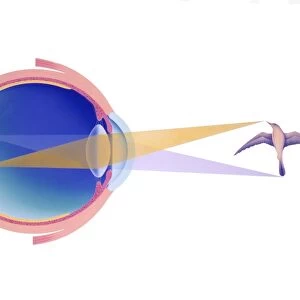
Eye anatomy. Artwork of a sagittal section showing the internal structure and anatomy of a human eye. The front of the eye is at right. Light entering the eye passes through the conjunctiva, cornea, and anterior chamber (green). After passing through the pupil, a hole surrounded by the iris that controls its size, the light passes through the lens (white). Here, it is focused on the retina, a light-sensitive layer rich in blood vessels lining the inside of the eye. The light triggers signals that are transmitted by the optic nerve to the brain. The optic nerve is at the back of the eye (lower left). Eye layers shown here include the sclera and choroid
Science Photo Library features Science and Medical images including photos and illustrations
Media ID 6319907
© CLAUS LUNAU/SCIENCE PHOTO LIBRARY
Aqueous Humour Choroid Conjunctiva Halved Iris Lens Ocular Ophthalmology Optic Nerve Pupil Retina Sagittal Sclera Sense Sensory Sight Vision Vitreous Humour Anterior Chamber Section Sectioned
MADE IN THE USA
Safe Shipping with 30 Day Money Back Guarantee
FREE PERSONALISATION*
We are proud to offer a range of customisation features including Personalised Captions, Color Filters and Picture Zoom Tools
SECURE PAYMENTS
We happily accept a wide range of payment options so you can pay for the things you need in the way that is most convenient for you
* Options may vary by product and licensing agreement. Zoomed Pictures can be adjusted in the Cart.