Home > Animals > Mammals > Muridae > Fortior
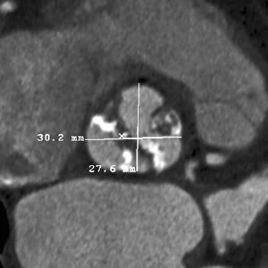
Balloon angioplasty, X-ray

![]()
Wall Art and Photo Gifts from Science Photo Library
Balloon angioplasty, X-ray
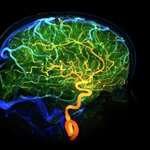
Balloon angioplasty. Coloured X-ray of the abdomen of a 45-year-old patient with a history of thrombosis (blood clots) in the inferior vena cava (vein), which have narrowed (stenosed), damaged and weakened the wall of the vein. This image shows the vein being repaired using a balloon angioplasty. A long thin tube with a deflated balloon at the tip (balloon catheter, centre left) is inserted into the blood vessel. When the catheter is within the narrowed region, the balloon is inflated to expand the vessel. A stent (wire mesh tube, lower centre) is then permanently implanted to hold the blood vessel open and reinforce the weakened wall
Science Photo Library features Science and Medical images including photos and illustrations
Media ID 9240557
© ZEPHYR/SCIENCE PHOTO LIBRARY
45 Year Old Angiogram Angioplasty Balloon Angioplasty Balloon Catheter Blood Flow Colored Device Diagnostic Imaging Forties Inferior Vena Cava Inserted Inserting Narrow Narrowed Procedure Radiography Radiological Radiology Stenosed Stenosis Stent Treated Treating Treatment Vascular Vascular Disease Xray Abnormal Blood Vessel Circulation Circulatory System Condition Deflated Disorder Unhealthy Vein
FEATURES IN THESE COLLECTIONS
> Animals
> Mammals
> Muridae
> Fortior
> Arts
> Minimalist artwork
> Monochrome artwork
> Fine art
> Arts
> Minimalist artwork
> Monochrome artwork
> Monochrome paintings
EDITORS COMMENTS
This print from Science Photo Library showcases the intricate procedure of balloon angioplasty, a revolutionary treatment for vascular diseases. The image depicts the abdomen of a 45-year-old patient with a history of thrombosis in the inferior vena cava, resulting in narrowed and weakened walls of the vein. In this technologically advanced medical intervention, a deflated balloon catheter is carefully inserted into the blood vessel through a long thin tube. Once positioned within the stenosed region, the balloon is inflated to expand and restore proper blood flow. To reinforce and maintain an open passage, a wire mesh tube called a stent is permanently implanted. The vivid colors bring attention to this life-saving technique that addresses circulatory disorders by treating narrow or abnormal blood vessels. Through radiography, healthcare professionals can precisely diagnose and monitor conditions affecting our biological systems. This monochrome image not only highlights the remarkable advancements in medical equipment but also emphasizes how technology intertwines with biology to improve human health. It serves as an educational tool for understanding anatomy while showcasing cutting-edge procedures that enhance our well-being. Science Photo Library continues its commitment to providing exceptional visual resources that contribute to scientific knowledge without mentioning any commercial use or specific company affiliation.
MADE IN THE USA
Safe Shipping with 30 Day Money Back Guarantee
FREE PERSONALISATION*
We are proud to offer a range of customisation features including Personalised Captions, Color Filters and Picture Zoom Tools
SECURE PAYMENTS
We happily accept a wide range of payment options so you can pay for the things you need in the way that is most convenient for you
* Options may vary by product and licensing agreement. Zoomed Pictures can be adjusted in the Cart.