Home > Popular Themes > Human Body
Ureteropelvic junction (UPJ) obstruction in the kidney
 obstruction in the kidney")
![]()
Wall Art and Photo Gifts from Stocktrek
Ureteropelvic junction (UPJ) obstruction in the kidney
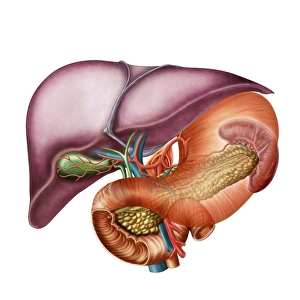
Ureteropelvic junction (UPJ) obstruction in the kidney. UPJ is defined as an obstruction of the flow of urine from the renal pelvis to the proximal ureter
Stocktrek Images specializes in Astronomy, Dinosaurs, Medical, Military Forces, Ocean Life, & Sci-Fi
Media ID 13013171
© Stocktrek Images
Anatomy Artery Biology Biomedical Illustrations Blood Flow Bulge Colored Background Cross Section Cutaway View Digestion Digestive System Filter Gland Healthcare Human Anatomy Human Body Parts Human Glands Human Kidneys Human Organs Interlobular Artery Internal Organs Kidney Major Calyx Medical Medicine Medulla Minor Calyx Nephrology Nephron Organ Physiology Renal Artery Renal Capsule Renal Circulation Renal Column Renal Hilum Renal Papilla Renal Pelvis Renal Pyramids Ureter Urinary System
FEATURES IN THESE COLLECTIONS
EDITORS COMMENTS
This print showcases the intricate details of a Ureteropelvic junction (UPJ) obstruction in the kidney. With its colored background and horizontal composition, this image provides a striking visual representation of this medical condition. The UPJ obstruction refers to the blockage that hinders urine flow from the renal pelvis to the proximal ureter. In this cross-section view, we can observe various components of the kidney's anatomy, including arteries, glands, and internal organs. The renal pyramids and medulla are clearly visible, highlighting their role in filtration and digestion processes. With its precise biomedical illustrations, this image serves as an invaluable resource for healthcare professionals specializing in nephrology or urology. It offers insights into renal circulation, blood flow patterns within human kidneys, and other aspects related to urinary system functioning. The cutaway view allows us to explore different structures such as minor calyxes, major calyxes, renal papilla, and interlobular arteries. Additionally, it highlights key features like the bulge at the ureteropelvic junction – a significant indicator of UPJ obstruction. Whether used for educational purposes or scientific research in physiology or biology fields; this visually appealing photograph is an excellent addition to any medical library. Its clarity and attention to detail make it a valuable tool for studying human anatomy while providing ample copy space for annotations or explanatory text.
MADE IN THE USA
Safe Shipping with 30 Day Money Back Guarantee
FREE PERSONALISATION*
We are proud to offer a range of customisation features including Personalised Captions, Color Filters and Picture Zoom Tools
SECURE PAYMENTS
We happily accept a wide range of payment options so you can pay for the things you need in the way that is most convenient for you
* Options may vary by product and licensing agreement. Zoomed Pictures can be adjusted in the Cart.